An ultrasound scan is one of the most important parts of a medical diagnostic and healthcare facility.
An ultrasound scan uses high-frequency sound waves to create images of the inside of the body.
Moreover, it is suitable for use during pregnancy as well.
These cans or sonography are safe as they use sound waves or echoes to make an image instead of using radiation.
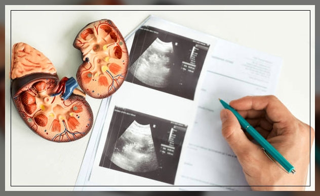
Furthermore, ultrasound scans help to evaluate fetal development and can also help detect problems in the liver, heart, kidney, or abdomen.
They can also assist in performing certain types of biopsies.
The image this can produce is a Sonogram.
It is important to note that these scans are safe and widely used around the world and often help to check the progress of the pregnancy.
However, doctors and radiologists can also use it for diagnosis and treatment.
Keep on reading.
Understanding Ultrasound Scan
The person who performs an ultrasound scan is a Sonographer, however, radiologists, cardiologists, or other specialists interpret the images.
Moreover, the sonographer often holds a transducer, a hand-held device, which looks like a wand.
They place it on the skin of the patient.
It is important to note that ultrasound is sound that travels through soft tissue and fluids.
However, it bounces back or echoes, off denser surfaces.
This is how it creates an image.

The term “ultrasound” refers to sound with a frequency that humans are unable to hear.
For diagnostic uses, the ultrasound is often between 2 and 18 megahertz, MHz.
Higher frequencies provide better quality images but are more readily absorbed by the skin and other tissue.
This is why they cannot penetrate as deeply as lower frequencies.
While lower frequencies tend to penetrate deeper, the image quality is inferior.
Learn more about Medical Diagnostic Imaging and blood test here.
How it captures Images?
Ultrasound tends to travel through the blood in the heart chamber for instance.
However, if it hits a heart valve, it will echo, or bounce back as a result.
It will travel through the gallbladder. And in case there are no gallstones, it will not bounce back.
However, if there are stones, they will bounce back from them.
Therefore, the denser the object the ultrasound hits, the more of the ultrasound will bounce back.
This bouncing back or echo gives the ultrasound image its feature.
While varying shades of grey reflect different densities.
Ultrasound Transducers
Doctors or sonographers will place the transducer or wand, normally on the surface of the body of the patient.
However, there are some kinds that can also place internally.
Some examples are:
- an endovaginal transducer for use in the vagina or transvaginal ultrasound
- an endorectal transducer for use in the rectum
- a transesophageal transducer that the doctor can pass down the throat of the patient for use in the esophagus
-
transrectal ultrasound
While some very small transducers can also be placed onto the end of a catheter and inserted into the blood vessels to examine the walls of the blood vessels.
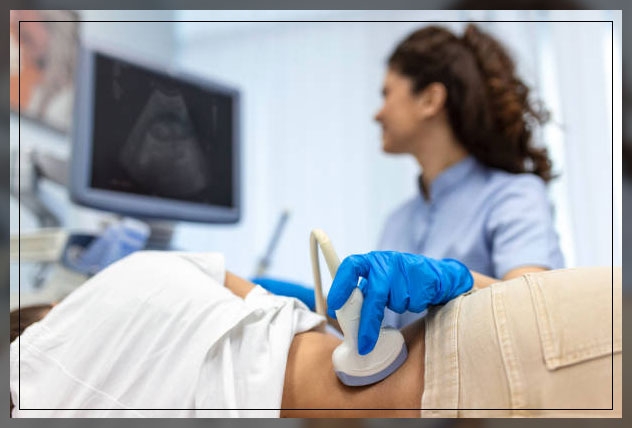
Procedure of Ultrasound
Before the exam, you will need to change into a hospital gown and you will most likely lie down on a table with a section of your body exposed.
An ultrasound technician or a sonographer will apply a special lubricating jelly to the skin.
This will help prevent friction so that the doctor can rub the ultrasound transducer on the skin.
The transducer looks like a wand or a microphone and the jelly helps to transmit the ultrasound waves.
The transducer sends high-frequency sound waves through the body of the patient, and the waves echo as they hit a dense object, like an organ or bone.
These echoes will then reflect into a computer.
Moreover, the sound waves are too high of a pitch for the human ear to hear and form a picture that doctors can interpret.
Depending on the area of examination, you may need to change positions so that the technician can have better excess.
After the procedure, the doctor will clean off the gel and the whole procedure often lasts for 30 minutes or so.
After the Exam
Following the exam, the doctor will review the images and check for any abnormalities and will call the patient to discuss the finding or to schedule a follow-up appointment.
In case they find anything abnormal, the patient may need to undergo other diagnostic techniques like:
- CT scan
- MRI
- A biopsy sample of tissue
However, it depends on the area they need to examine.
Therefore, if the doctor is able to make a diagnosis of the condition of the patient depending on the ultrasound, they can begin the treatment immediately.
Uses of Ultrasound Scan
In most cases, doctors use ultrasound scans for diagnosis, treatment, and guidance during certain procedures like biopsies.
It can also help to examine internal organs like the liver and kidneys, the pancreas, the thyroid glands, the testes, ovaries, and others.
Moreover, an ultrasound scan can also help to reveal whether a lump in the body of the patient is a tumor or not.
This can be cancerous or a fluid-filled cyst.
Furthermore, it can help diagnose issues with soft tissues, muscles, blood vessels, tendons, and joints.
Certain other issues it can help to investigate are:
- frozen shoulder
- tennis elbow
- carpel tunnel syndrome, and others
Learn more about Radiology Tech here.
Ultrasound Scan for Circulatory Problems
Doppler ultrasound can assess the flow of blood in a vessel or blood pressure.
It can also find the speed of the blood flow and any obstruction.
For instance, an echocardiogram, or ECG is an example of a Doppler ultrasound.
Doctors can use it to create images of the cardiovascular system and to measure blood flow and cardiac tissue movements at specific points.
Moreover, a Doppler ultrasound can help to assess the function and state of cardiac valve areas, any abnormalities in the heart, valvular regurgitation, or blood leaking from valves.
It can also show how well the heart is pumping out blood.

Doctors can also use it to:
- examine the walls of blood vessels
- check for DVT or an aneurysm
- check for fetal heart and heartbeat
- assess for blockages or narrowing of arteries.
On the other hand, a carotid duplex is a form of carotid ultrasonography that can also include a Doppler ultrasound.
This can help reveal how blood cells move through the carotid arteries.
Use of Ultrasound in Anesthesiology
One of the important uses of ultrasound scans is its use by anesthetics to guide a needle with an anesthetic solution near nerves.
An ultrasound can take place at the doctor’s office, at an outpatient clinic, or in the hospital.
Moreover, in most cases, it takes about 20 to 60 minutes and is not normally painful and there is no noise as well.
Also, patients do not need to prepare specially for an ultrasound, however, they may wish to wear loose-fitting and comfortable clothing.
In case the liver or gallbladder is affected, the patient may need to fast or eat nothing, for at least several hours before the procedure takes place.
While for a scan during pregnancy, and especially during early pregnancy, the patient will need to drink plenty of water.
Furthermore, they should also try to avoid urinating for some time before the test.
When the bladder is full, the scan will produce a better image of the uterus.
The scan often takes place in the radiology department of a hospital and a doctor or a specially-trained sonographer will carry out the test.
External Ultrasound Scan
During an external ultrasound, the sonographer will place a lubricating gel onto the skin of the patient and place a transducer over the lubricated skin.
They will then move the transducer over the part of the body that needs to be examined.
Certain examples are ultrasound examinations of the heart of the patient or a fetus in the uterus.
Moreover, the patient should not feel any discomfort or pain, while they will just feel the transducer over the skin.
However, during pregnancy, there may be slight discomfort because of the full bladder.
Internal Ultrasound Scan
In case doctors need to evaluate internal reproductive organs or urinary systems, they will place the transducer in the rectum of a main or the vagina of a woman.
For instance, they evaluate some parts of the digestive system, the esophagus, the chest lymph nodes, or the stomach, they may use an endoscope.
A light and an ultrasound device are attached to the end of the endoscope, which they will insert into the body of the patient, often through the mouth.
However, before the procedure, the doctor may give medications to reduce any pain.
Moreover, internal ultrasounds are less comfortable than external ones.
While there is a slight risk of internal bleeding, a patient should seek immediate medical advice.
Safety
It is important to note that most types of ultrasound are noninvasive and they involve no ionizing radiation exposure.
Many experts believe this procedure to be very safe.
However, since the long-term risks are not established, unnecessary ‘keepsake’ scans during pregnancy are not encouraged.
Moreover, ultrasound during pregnancy is recommended only when medically needed.
In case anyone is allergic to latex, they should inform their doctor so that they will not use a latex-covered probe.
Final Thoughts
An ultrasound is a medical test that uses high-frequency sound waves bounce to capture live images from inside the body of the patient.
The technology is just like that of sonar and radar. An ultrasound allows the doctor to see problems with organs, vessels, and tissues that need to make an incision.
Unlike other imaging scans, ultrasound uses no radiation, and for this, they are often used for viewing a developing fetus during pregnancy.