CT scans or CAT scans are an important part of any medical health care facility.
Doctors and healthcare professionals have years of training in their respective fields.
However, there are still a lot of things they are unable to diagnose simply by looking at or listening to your body.
Certain medical conditions need a deeper look, usually at the tissues, blood vessels, and bones inside your body.
Though X-rays and ultrasounds can provide some information, however when they need a more detailed view, a computer tomography, or CT scan is often the next step.
A CT scan combines a series of X-ray images that it takes from different angles around the body of the patient.
And uses computer processing to create cross-sectional images, or slices of the bones, blood vessels, and soft tissues.
Moreover, it provides more detailed information than a plain X-ray.
Keep on reading to learn more about CT scans.
CT Scans
A CT scanner emits a series of narrow beans through the body of the patient as it moves through an arm.
This is different from an X-ray machine.
An X-ray machine sends just one radiation beam, while a CT scan prodcues more detailed final pictures than an X-ray image.
Moreover, the X-ray detector in the CT scanner can see hundreds of different levels of density and it can see tissues within a solid organ.
This data is then transmitted to a computer.
There it builds up a 3-D cross-sectional picture of the part of the body and displays it on the screen.
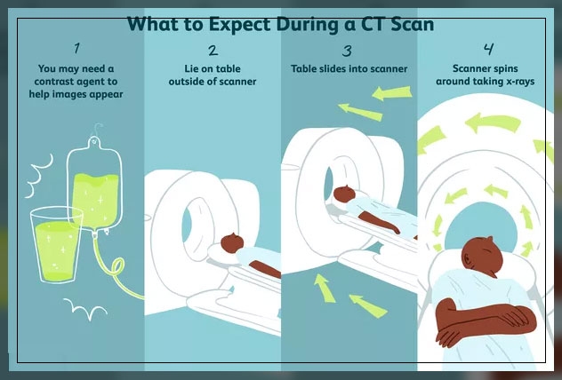
In some cases, a contrast dye can also be used.
It helps to show certain structures more clearly.

For instance, if a 3-D image of the abdomen is required, the patient may have to drink barium mean.
This barium appears white on the scan as it travels through the digestive system of the patients.
However, if the detailed images lower down the body are required, like that of the rectum, the doctor may give barium enema to the patient.
In case they need blood vessel images a contrast agent in the veins can help.
Furthermore, the accuracy and speed of the CT scan are improved with the application of spiral CT, a relatively new technology.
The beam takes a spiral path during the scanning and gathers continuous data with no gaps between images.
CT is a useful tool for assisting diagnosis in medicine, however, it is also a source of ionizing radiation and can potentially cause cancer.
Therefore, the National Cancer Institute advises patients to discuss the risks and benefits of CT scans with their doctor.
Use of Contrast Dye
The images a CT scan provides appear in shades of blacks and greys.
It can sometimes be difficult for even a trained eye to differentiate one tissue type from another in some conditions.
Therefore, doctors or radiologists may use contrast dye that contains barium or iodine.
Patients can either take them orally or intravenously, through the vein.
These dyes help to increase the contrast levels and resolution of the final images a CT prodcues for a more exact diagnosis.
However, there a certain risks that come with using contrast dyes.
For instance, there is a higher chance of allergic reaction to the dye and they are also not beneficial for your kidneys.
Stills, every CT scan exposes the patient to a certain level of radiation, and CT with contrast dye may produce better results than one without.
It can also help to prevent the need for repeated scans.
The following is the comparison of when doctors can use CT can with or without a contrast dye:
| With contrast | Without contrast |
|---|---|
| acute appendicitis | acute stroke |
| staging cancer | closed head injuries |
| diverticulitis | lung disease |
| inflammatory bowel disease | tissue swelling or injury in your arms or legs |
| pancreatitis | kidney stones |
Uses of CT Scans
These scans are useful to obtain the images of:
- soft tissues
- the pelvis
- blood vessels
- lungs
- brain
- abdomen
- bones
Many doctors and healthcare professionals consider CT a way of diagnosing different types of cancer, like liver, lung, and pancreatic cancer.
The image allows them to confirm the presence and location of a tumor, its size, and how much it is affecting the nearby tissues.
Moreover, the scan of the head provides important information about the brain, for instance.
If there is bleeding, swelling of the arteries, or a tumor.
A CT scan can help reveal a tumor in the abdomen, and any swelling or inflammation in nearby internal organs.
Furthermore, it can also show any laceration of the spleen, kidneys, or liver.
As CT scans detect abnormal tissue, it is useful for planning areas for radiotherapy and biopsies and can provide valuable data on blood flow, heart disease, and other vascular conditions.
Also, it can help the doctor assess bone diseases, bone density, and the state of the spine of the patient.
It can also provide important data about injuries to the hands, feet, and other skeletal structures of the patient.
Even small bones are visible on CT scans.
Learn more about Medical Diagnostic Imaging here.
CT vs. MRI
The major differences between CT and MRI are:
- A CT scan uses X-rays, however, MRI uses magnets and radio waves
- Unlike an MRI, CT scans do not show tendons and ligaments
- MRI is better for the examination of the spinal cord

- A CT scan is better suited to conditions like cancer, pneumonia, abnormal chest x-rays, and bleeding of the brain, especially after an injury
- A brain tumor is more likely to be visible on MRI
- CT scans how organ tear and organ injury more quickly, so it may be more suitable for trauma cases
- In the case of broken bones and vertebrae, CT scans show more visible images
- CT provides a better image of lungs and organs in the chest cavity between the lungs
Learn more about X-ray Machine: Overview here.
On the day of the Procedure
In some cases, patients may need to abstain from food and possibly drink for a certain period before the scan.
In most medical and diagnostic facilities, the patient will need to undress, usually down to the underwear, and put on the gown that the health canter will provide.
Moreover, it is important that the patient is not wearing jewelry.
If the hospital does not provide a gown, the patient should wear loose-fitting clothes free of metal objects, buttons and zippers.
While some patients may have to drink a contrast dye, or the doctor may give the dye as an enema or injection.
This will help to improve the picture of some blood vessels or tissues.
However, any patient who has an allergy to contrast material should tell the doctor beforehand.
Some medications can help to reduce allergic reactions to contrast material.
As metal tends to interfere with the workings of the CT scanner, the patient will have to remove any jewelry and metal fastenings.
In case the patient is wearing glasses, watches, hairpins, hearing aids, denturs, bras with underwire, ‘antimicrobial’ clothese with silver technology, nicotine patches, or other medication patches, they should remove them.
During and after CT Scans
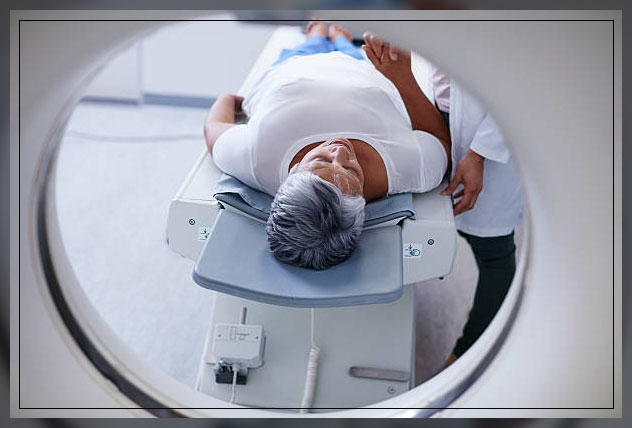
During the scan, the patient will need to lie down on a motorized examination table that will slide into a doughnut-shaped CT scanner machine.
In most cases, the patient will lie down back, facing up, however, in some cases, they may need to lie facedown or sideways.
After one x-ray picture, the couch will move slightly, and then the machine will take another image, and so on.
Moreover, the patient needs to stay or lie very still and hold your breath in order to get the best results.
During the scan, everyone except for the patient will leave the room.
An intercom will enable two-way communication between the padiographer, or doctor, and the patient.
However, if the patient is a child, a parent or an adult mayallowedowes to stand or sit nearby, but they will havelearnlear a lead apron to prevent any radiation exposure.
Once the CT scan is over, the radiologist will see the images for examination.
A radiologist is a doctor who specializes in the diagnosis and treating conditions using imaging techniques like CT scans, and X-rays.
Your doctor will follow up with you to explain the results afterward.
Risks of CT Scans
A CT scan involves a small, targeted dose of radiation, and these levels of radiation therapy, have not proven to be harmful.
Even in people who undergo a number of scans.
The chance of developing cancer as a result of getting a CT scan is thought to be less than 1 in 2,000, according to the Society of North America.
Moreover, the amount of radiation in this scan, according to estimates is around the same as a person who has exposure to a space between several months and several years of natural exposure in the environment.
The doctor will give you a scan if there is a clear medical reason to do so.
The result can lead to treatment for conditions that can otherwise be seriuos.
When the doctor or healthcare professional takes the decision to perform a CT scan, they will make sure that the benefits outweigh any risk.
Though problems can arise from exposure to radiation including cancer and thyroid, it is rare.
Furthermore, these are extremely rare in adults and also unlikely to be in children.
However, they tend to be more susceptible to the effects of radiation.
This does not mean that a person will have health issues.
But any CT scan should be noted on the medical records of the child.
In some cases, only a CT scan can show the required results and for some conditions, an ultrasound or MRI may also be possible.
Learn more about Radiology Center Near Me here.
FAQs
Can a patient have a CT scan if pregnant?
Any woman who is suspecting that she is pregnant should tell the doctor beforehand, as there is a risk that an x-ray can harm the fetus.
Citing the American College of Radiology, the American Pregnancy Association, APA, points out that:
“No single diagnostic X-ray has a radiation dose significant enough to cause adverse effects in a developing embryo or fetus”.
However, APA also notes that CT scans are not recommended for pregnant women unless the benefits outweigh the risk.
Can a nursing mother have a CT scan?
In case a lactating or breastfeeding mother needs an iodinated intravenous dye for contrast, she should avoid breastfeeding for at least 24 hours, according to the Radiology Society of North America.
This is because the dye may pass into breast milk.
What can you do if the patient is claustrophobic?
In case the patient is claustrophobic, they should tell the doctor or radiographer beforehand.
The doctor or radiologist may give an injection or tablet to the patient to calm them down before the scan.