
Medical Diagnostic Imaging is a crucial part of any healthcare facility.
Diagnostic health imaging technology has transformed healthcare over the years and now allows for earlier diagnosis of medical conditions.
Moreover, it also helps to reduce the need for needless invasive exploratory processes and creates better patient outcomes.
Medical diagnostic imaging also offers comprehensive diagnostic imaging services.
It is important to note that it is a common technique that helps to visualize physical diagnosis in the clinical community.
Certain techniques that fall under the medical imaging umbrella are radiology, magnetic resonance imaging, MRI, computed tomography, CT, fluoroscopy, ultrasound, echocardiography, and nuclear medicine, like PET.
For diagnostic purposes, these systems help to image the body in order to obtain a diagnosis and determine future care.
Rapid advancements in technology and changes in healthcare reforms help to take a mindful approach in order to end up with the correct system for your facility.
Keep on reading to learn more about it.
Medical Diagnostic Imaging and its Uses
This diagnostic imaging describes different techniques for viewing the insides of the body.
Moreover, they help to figure out the causes of an illness or injury and confirm the diagnosis.
Doctors and other healthcare facility members also use it to see how well the body of their patients responds to treatment for a fracture or illness.
Diagnostic imaging also allows the physician to view the inside of the body of the patient to help find any indications of a health condition.
Furthermore, some machines and methods can help produce pictures of the activities and structures inside the body.
Thus, your doctor can decide which medical imaging tests to use based on the part of the body they are evaluating and the symptoms.
Many imaging tests are non-invasive, easy, and painless.
Some will need patients to remain inside the machine for a long time.
However, it can get a little uncomfortable.
Some tests involve a small amount of radiation exposure as well.
While for other imaging tests, doctors may insert a small camera attached to a thin, long tube into the body.
This device is a scope. Doctors can then move the scope through a bodily opening or passageway to view the insides of an organ, like lungs, heart, or colon.
However, patients may need anesthesia for these procedures.
Let’s discuss the types of medical imaging diagnostics as follows:
MRI Scans
MRIs do not use radiation, however, make use of a powerful magnet to obtain an image of the body of the patients.
There are four types of MRI machines:
Ture Open A true open MRI design is open on all sides.
And removes a lot of discomfort for individuals who are claustrophobic in a regular MRI machine.
Closed This is a closed machine or a traditional tube machine.
Moreover, it allows the patient to lie down and go inside for the images.

3T The “T” means Tesla and is a unit of measurements the technologists often uses to qualify the magnetic field strength.
Additionally, the 3T is the most innovative and advanced test of all MRIs that are used today.
It is important to note that it consists of a closed design like a traditional MRI.
However, it takes less time to perform the uses using the 3T MRI scan the images are detailed and high resolution.
Thus enabling the radiologists to determine where the patients have a benign or more severe medical condition.
Wide Bore Also called an “open MRI” this machine resembles a closed MRI, however, with a wider opening.
Learn more about Infection Control and Prevention Guidelines here.
Uses of MRI Scans
As a doctor, you may recommend an MRI scan for a number of reasons.
It provides an incredibly detailed image or look inside the body and can help to examine things like:
Spinal cord and brain abnormalities, cysts, tumors, other bodily irregularities, joint abnormalities, and injuries.
Moreover, you may recommend it for:
- breast tissues to screen for cancer
- the pelvic region of a female to identify issues like fibrosis and endometriosis
- suspect uterine anomolies
- abdominal or liver disease
It is important to note that MRI technology is continually expanding and often takes about 30 to 60 minutes to complete.
You may choose to use contrast or fluid injection into the vein of the patient to make certain details clear in the generated images.
This step, however, can make the exam last longer.
Learn more about Medical Items: Guide here.
MRA Scans
MRA stands for Magnetic resonance angiogram.
This test helps to provide very detailed images of the blood vessels in the body. MRA scans are a form of MRIs.
Moreover, these machines provide information that CT scans, ultrasounds, or X-rays are unable to provide as they use radio wave technology and a magnetic field.
As a doctor, you may use them on the legs, neck, brain, and kidneys in order to collect information about the condition of blood flow and vessel walls.
Doctors use MRAs to also look for calcium deposits, aneurysms, and clots within the blood vessels.
In some cases, however, a contrast dye can help to get a better definition of the images of the blood vessels.
Furthermore, MRA offers to provide definite benefits to both you and your patients.
The benefits of using MRA are:
- they do not make use of radiation like CT scans, X-rays
- are non-invasive
- can help detect information that x-rays, ultrasounds, and CT scans may miss
- detect issues with blood vessels that lead to a reduction in the blood flow
Learn more about Medical Imaging here.
Uses of MRA scans
An MRA scan is a helpful diagnostic tool.
Its main purpose is to help find issues that may exist within the blood vessels.
As a doctor, you may use them for detecting calcium deposits, aneurysms, or clots in blood vessels, and find existing blood vessel narrowing.
Moreover, you may use it to:
- identify abnormalities in the blood vessels of the brain like congenital disabilities and inflammation
- defining blood supply to the vascular tumors of the brain
- help evaluate stroke patients, etc.
Learn more about Medical Equipment Suppliers in Dubai here.
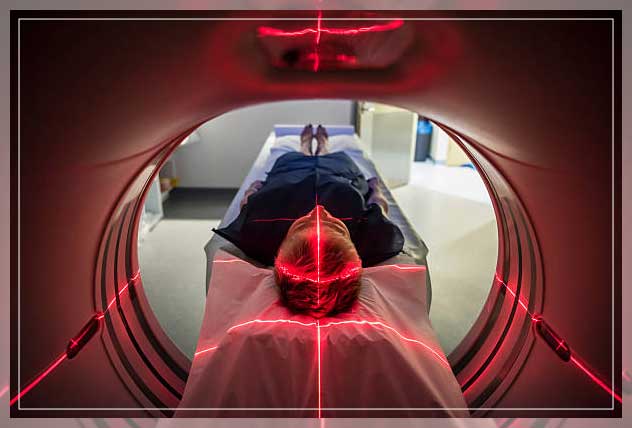
CT Scans
Physicians can refer to a CT scan as a “CAT Scan” as well.
This can or test combines a string of X-ray scans or images that it takes from various angles.
Computer software then generates cross-sectional images or slices of blood vessels and soft tissues inside the body.
CT also offers more thorough images than standard X-rays.
Their frequent use help to quickly examine individuals who have internal injuries from trauma.
Doctors can use these scans to evaluate the spine, brain abdomen, neck, and chest of the patients.
Moreover, they provide clear images of both hard and soft tissues.
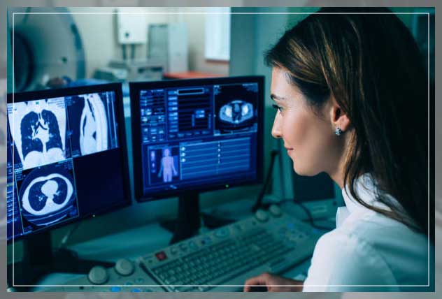
The images that come from a CT scan also allow doctors to make a quick medical decision.
It is important to note that CT scans are performed at both imaging centers and hospitals.
They help physicians find injuries and diseases that can be previously found with surgery or autopsy.
Uses of CT scan
While CT scans use low doses of radiation, they are relatively non-invasive and safe.
Furthermore, these scans are useful in a number of medical situations where diagnostic imagery is important.
They can even assess slight abnormalities in soft tissues like the brain as well as other organs.
Doctors can use the images when patients have certain symptoms like dizziness or pain.
They are even useful in examining the spread of conditions like care.
Depending on where the technologists direct the CT Scan in your body, there are different uses for it.
These are:
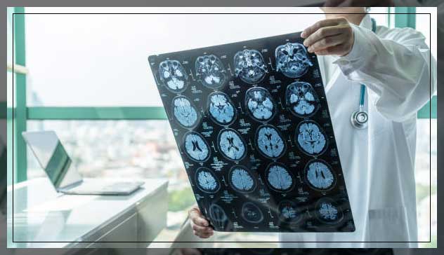
Brain or head CT Scan to check for stroke, bleeds, masses, and other abnormalities and examine the skull.
Chest CT Scan to provide further insight into abnormalities after a standard X-ray
Neck CT Scan to look for enlarged glands or lymph nodes and study lumps
Spine CT Scans help to detect spine issues like spinal cord narrowing, a herniated disc, or fractures
Sinus CT Scans helps to detect and diagnose obstructions or sinus disease
Pelvic or Abdominal CT Scans help to check organs in this area and diagnose unexplained pain in the abdomen.
Ultrasounds
Another important medical diagnostic imaging includes Ultrasound.
Doctors also refer them to as ‘sonography’ which is a safe imaging method that helps creates images of the insides of the body.
It does not use radiation, however, high-frequency waves.
As a result, it is a safe procedure even during pregnancy.
Moreover, the ultrasound images are in real-time and show the structure and movement of internal organs as well as the blood flow through vessels.
During an ultrasound, a sonographer will hold a transducer, a handled device over the skin.
In some cases, they may place it internally.
This machine uses sound waves traveling through soft tissues and fluids and as it hits denser surfaces, it echoes or bounces back.
Thus, resulting in images. More ultrasound echoes back when the object is denser.
Learn more about Dental Implants here.
Uses of Ultrasound
As a doctor, you can diagnose a large variety of health conditions with this medical diagnostic imaging.
Furthermore, the images it creates help you to come up with effective treatment plants.
In cases patients have symptoms like swelling, infection, or pain, you can also suggest an ultrasound to determine the cause.
However, in some cases, you may use these machines to assist anesthesiologists during surgical procedures when they are guiding needles near nerves.
In most cases, ultrasounds are tools that allow you to examine problems related to the abdomen, circulation, urology, obstetrics, newborn care, and even musculoskeletal conditions.
Some common body parts physicians use ultrasound includes:
- heart
- joints
- uterus
Moreover, they also use it for the bladder, kidneys, and blood vessels.
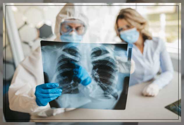
X-ray and Mammography
One of the most common medical diagnostic imaging includes X-rays.
Doctors use them to view the insides of the body.
X-ray machines produce a high-energy beam that dense tissues and bones are unable to sort, however, tends to pass through other areas.
This process, results in an image, allowing you to see if the patient is suffering issues ranging from an injury to bone issues.
On the other hand, mammography is a type of X-ray image of breasts.
They help to check for breast cancer signs like small lumps on the breasts you are otherwise unable to feel through the use of a low-dose x-ray.
Moreover, they also show breast tissue changes that can potentially be a sign of early-stage breast cancer.
A radiologist uses digital mammography to identify and diagnose cancer nodules that older systems are unable to detect.
Moreover, mammograms are the best way to detect breast cancer.
It also has a number of benefits that includes:
- detecting breast cancer early on
- reduces your risk of dying because of cancer by 30%
- treatment early on so that you do not have to resort to mastectomy
Bone Density Scans
A bone density scan is an indirect scan you can use to determine whether or not your patient has osteoporosis.
This procedure is also referred to as “bone mineral density testing” as it measures the amount of bone material you have per square centimeter in your bone.
Osteoporosis is a condition that can make your bones fragile and susceptible to fractures.
With the help of bone density scans that use x-ray equipment to measure bone minerals and calcium in a small bone, doctors can make a diagnosis.
Often, you will have this can on your hip, spine, or forearm.
Bones tend to be denser if you have higher bone mineral content.
This means that they are less likely to break.
However, the bones are ore at risk of fractures if they have low bone mineral content, which also indicates osteoporosis.
Prior to using this scan, the only way you could diagnoses osteoporosis was if the bone breaks.
However, at this point, the bones are already weak and in a poor state.
it is important to note that anyone can develop this condition, however, women are more prone to developing it.
You can suggest this can if:
- there are fragile bones susceptible to fractures
- reduction in height of a minimum of an inch and a half
- lower sex hormone levels
- need for certain medications that can interfere with the rebuilding process of bones like steroids
- need for anti-rejection medication after a transplant
Arthrogram and Myelogram
When the joints do not function as they should, stops the movement ability, and makes daily tasks harder, you may need an arthrogram.
An arthrogram is one of the different types of medical diagnostic imaging that helps to diagnose joint problems.
It is helpful when other types of imaging might not be able to detect.
Also referred to as ‘arthrography’ these consist of different images that use x-ray fluoroscopy, CT scans, or an MRI specifically of the joints.
Moreover, the radiologist will inject the joint with a contrast dye like iodine before the procedure begins.
They will use a fluoroscope to guide the injection placement into the joints of the patients.
This dye will coat the joints’ structure linings that make them look white on the images and highlights any issues so that you can evaluate the function of the joints.
On the other hand, a myelogram is helpful when you need specific imaging of the spinal canal.
This includes issues like spinal tissues, spinal cord, ad surrounding nerves.
A myelogram is an exam during which the technologist will insert a contrast dye into the spinal cord space while using fluoroscopy to take moving x-ray images.
As this dye flows through spaces, you can examine the area of abnormalities, including:
- tumors
- infection
- inflammation
A CT scan often follows myelogram procedures that he’s to better define any possible issues.
Along with CT technology, a myelogram can help to give more detailed information that you can get along with x-rays alone.
Schedule an Appointment
Understanding what your healthcare facility needs is an important part to make sure you provide all the important medical diagnostic imaging facilities.
We at Engiomed have top-notch quality products for your healthcare facility o diagnostic centers. Book an appointment today to find out more about us.


Comments