A PET scan or Positron Emission Tomography is an imaging scan that helps to reveal the metabolic or biochemical function of the tissues and organs.
Positron emission tomography, PET scan uses a radioactive drug or tracer that helps to show both normal and abnormal metabolic activity.
Moreover, it can help to detect the abnormal metabolism of the tracer in diseases before the disease shows up on other imaging tests.
These include computerized tomography, CT, magnetic resonance imaging, and MRI.
It is important to note that the doctor will inject the tracer into the vein of the patient within the arm or hand of the patient.
This tracer will then collect into areas of the body of the patient that have higher levels of metabolic or biochemical activity.
Furthermore, this will help pinpoint the location of the disease.
PET scans are often combined with CT or MRI and are called PET-CT or PET-MRI scans.
Keep on reading.
Why do doctors Perform PET scans?
The doctor can order an emission tomography PET scan to inspect the blood flow in the body of the patient, the oxygen intake, or the metabolism of the organs and tissues.
PET scanners help to show problems at the cellular level, giving the doctor the best view of complex system diseases.
Moreover, doctors commonly use PET scans to detect:
- cancer
- heart problems
- brain disorders including problems with the central nervous system, CNS
Cancer
It is important to note that cancer cells tend to have a higher metabolic rate than noncancerous cells.
Because of the high level of chemical activity, cancer cells often show up as bright spots on PET scans.

For this very reason, doctors will use PET scans for both detecting cancer and:
- seeing if cancer has spread
- seeing if cancer treatment like chemotherapy is working or not
- checking for cancer recurrence
However, it is important for the doctor to read these scans carefully and explain them.
As it is often possible for noncancerous conditions to look like cancer on the scan and it is also common for solid tumors to fail to appear on PET scans.
Heart Problems
PET scans can show or reveal areas of decreased blood flow in the heart, which occurs when healthy heart tissue tends to take in more of the tracer than unhealthy tissue or tissue with a decrease in blood flow.
Moreover, different colors and degrees of brightness on the scan show different levels of tissue functions.
With the help of this information, the doctor can decide on how to move forward with treatment.
Brain Disorders
Glucose is the main fuel of the brain, and during PET scans, tracers “attach” themselves to compounds like glucose.
By detecting radioactive glucose or nuclear medicine, a PET scan can help to show areas in which areas of the brain are using glucose at the highest rates.
When a specialist interprets the scan, they can see how the brain is working and can check for any irregularities.
Furthermore, ETC scans can help to diagnose and manage a number of CNS disorders like:
- Alzheimer’s disease
- epilepsy
- depression
- head trauma
- Parkinson’s disease
Learn more about Medical Imaging here.
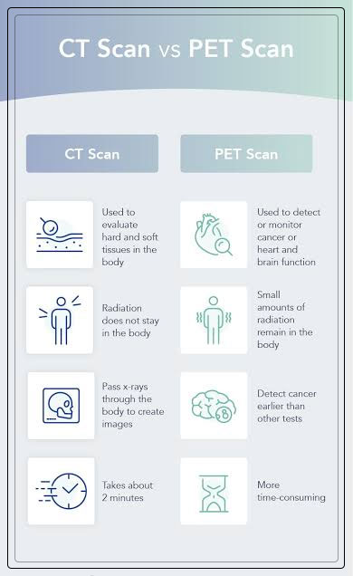
Comparing PET scans to other Tests
PET scans can help to show metabolic changes that occur at the cellular level in an organ or tissue of the patient.
This is important as diseases often begin at the cellular level.
CT scans and MRIs cannot, however, reveal such problems at the cellular level.
Moreover, PET scans can help detect very early changes in your cells.
On the other hand, CT scans, and MRIs can only detect changes later, as a disease alters the structure of the organs or tissues of the patient.
Detecting illness at the cellular level gives the doctor the best view of complex system diseases.
These include:
- coronary artery disease, CAD
- brain tumors
- memory disorders
- seizure disorders
In some cases, doctors can use PET scans along with other tests to get a clearer picture of the inside of the body.
This is also known as PET/CT or a PET/MRI scan.
In such cases, the tests a doctor performs will take place at the same time.
- On its own, a CT scan uses special X-ray equipment to produce pictures of the inside of the body.
- However, MRI scans use magnetic fields and radio frequency pulses to create images of internal structures like organs, soft tissues, and bones.
When a doctor performs either of the above in conjunction with a PET scan, the result is an Image Fusion.
The computer will combine the images from the two scans to create a three-dimensional image.
It then provides more information and allows for a more precise diagnosis.
Additionally, Gallium scans are just like PET scans
They involve injecting gallium citrate, a radioactive tracer into the body of the patient.
Gallium scans are a multiday process and doctors will perform 1 to 3 days after administrating the tracer.
It is important to note that these can are not commonly performed for the detection of cancer.
Though some forms of the gallium scan can be combined with newer tests like PET scan.
Learn more about Dental X-Ray: All You Need to Know here.
Are there any Risks?
The PET scan uses radioactive tracers, however, the exposure to harmful radiation is minimal.
The amount of radiation in the tracer is small, so the risks to the body of the patient are low.
Moreover, doctors and experts consider tracers radiopharmaceuticals.
And must meet Food and Drug Administration, FDA standards for both safety and performance.
Still, it is a good idea that the patient should discuss the possible risks with the doctor.
It is important to note that the risks of the test are also minimal in comparison to how beneficial the results can be to diagnosing seriuos medical conditions.
Furthermore, the tracer is essentially glucose with radioactive components attached to it which makes it very easy for the body to eliminate the tracer naturally after the test.
It will do so, even if the patient has a history of kidney disease or diabetes.
Patients with Allergies and Other Health Conditions
It is possible for a patient to have an allergic reaction to the tracer.
People who are allergic to iodine, aspartame, or saccharin should inform the doctor before.
Moreover, those who cannot have iodine will receive a tracer made of diluted barium sweetened with saccharin.

While those most likely to have an allergic reaction to the iodine tracer include people with:
- a history of allergic reactions to PET scans
- allergies
- asthma
- heart disease
- dehydration
- the blood cell disorders like sickle cell anemia, polycythemia vera, and multiple myeloma
- kidney disease
- a drug regimen including beta-blockers, non-steroidal anti-inflammatory drugs, NSAIDs, or interleukin-2, IL-2
Risks for people who are Pregnant, and Other Risks
It is important to note that doctors and experts do not consider radiation safe for developing fetuses.
Therefore, if the patient is pregnant, or thinks they are, they should not get a PET scan.
Moreover, if a patient is getting a PET/CT scan, they will need an additional tracer.
This can be harmful to people with kidney disease or those who have elevated creatinine levels from medications they are already taking.
While other risks of the test include discomfort if the patient is claustrophobic, or uncomfortable with needles.
The injection can also lead to symptoms like bleeding, bruising, or swelling.
Learn more about Blood Test: All You Need to Know here.
Preparing for PET Scans
The doctor will provide complete instructions on how to prepare for a PET scan.
The patient needs to talk to the doctor about any prescription, over-the-counter, OTC, or supplemental medications they are taking.
A few days before
Your doctor will ask the patient to avoid strenuous physical activity like exercises and deep-tissue massages in the 24 to 48 hours before the test.
The Day before the Test
In the 24-hour hours before the appointment of the patient, the doctor will ask to stick to a low carbohydrate, no sugar diet.
Moreover, food and beverages a patient should avoid are cereal, pasta, bread, rice, milk, and yogurt whether airy or nondairy, fruits and fruit juices, alcohol, caffeinated beverages, and candy including gum and mints.
Foods a patient can eat are meat, tofu, nuts, and nonstarchy vegetables like:
- carrot
- asparagus
- broccoli
- salad greens
- quash and more
Hours Before
If the patient receives anesthesia for the procedure, it is important to avoid drinking or eating anything the entire morning before the PET scan.
However, a patient can drink a few sips of water if they need to take any medication.
In case they are not receiving anesthesia, they should refrain from eating anything for 6 hours before the scan.
Remember that they should also avoid chewing gum or sucking on hard candy, cough drops, or mints.
A patient will be able to drink water and take medications as recommended by the doctor.
Moreover, the doctors will ask the patient to change into the hospital gown.
This is because metal can interfere with the testing equipment, so the patient will need to remove any jewelry they may be wearing, including body-piercing jewelry.
If they are undergoing PET?CT scans, medical devices like pacemakers, and artificial hips will not affect the results.
However, a patient cannot undergo a PET/MRI scan with unapproved medical devices or metal implants.
Other Considerations
The patient needs to tell the doctor about any medical condition they may have.
These include:
If a patient is pregnant or believes they could be pregnant, they should inform the doctor.
This is because this test is unsafe for the baby.
Breastfeeding Mothers: A patient may need to pump and store the breast milk 24 hours before the procedure.
Moreover, a patient will not be able to breastfeed for 24 hours after the test.
Diabetes: If a patient has diabetes, they will get special instructions for the test preparation as fasting beforehand can affect the blood sugar levels.
Furthermore, the patients will likely be asked to hold the normal dose of insulin and eat a light meal 4 hours before the scan is scheduled.
Performing a PET Scan
Before the scan, the doctor will inject the patient with a tracer through a vein in the arm, through the solution they will drink, or in a gas they inhale.
The body of the patient will need time to absorb the tracer, so they will need to wait an hour before the scan begins.
How long it takes for the body to fully absorb a tracer depends on the area of the body the doctor needs to scan.
While a patient waits, they will want to limit movement, relax, and try to stay warm.
Moreover, if they are undergoing a brain scan, they will want to avoid watching television, music and reading.
Next, they will undergo the scan, which can last anywhere from 30 to 45 minutes.

This will involve lying on a narrow table attached to a PET machine that looks like a giant letter “O”.
The table will glide slowly into the machine so that it can conduct the scan.
However, if a patient is undergoing multiple scans, they can add additional time, up to about 3 hours.
A patient will need to lie still during the scan, the technician will let them know when they need to remain still, and may be asked to hold their breath for several seconds.
Also, they will hear buzzing and clicking noises during the test.
When all the necessary images have been recorded, the patient will slide out of the machine, and the test is complete.
What happens after the Test?
After the test, the patient can go about their day unless the doctor gives other instructions.
However, as the radioactive material can remain in the body of the patient for a few hours to days, they should limit the contact with both pregnant people and infants during this time.
Moreover, they should drink plenty of fluids after the test to help flush the tracer out of the system.
Meanwhile, a trained specialist will interpret the positron emission tomography PET scan images and share the information with the doctor.
The results are usually ready for the doctor within a few days and will go over the results with the patient at the follow-up appointment.