Magnetic resonance imaging, MRI scans are common procedures around the world.
MRI scans use a strong magnetic field and radio waves to create detailed images of organs and tissues within your body.
Since its invention, doctors and researchers continue to refine MRI techniques to assist in medical procedures and research.
The development of MRI revolutionized medicine.
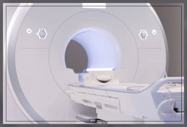
Most MRI machines are large, tube-shaped magnets.
When patients lie inside an MRI machine, the magnetic field temporarily realigns water molecules in the body.
Moreover, radio waves cause these aligned atoms to produce faint signals.
These are then used to create cross-sectional MRI images like slices in a loaf of bread.
Furthermore, the MRI machine can help create 3D images that doctors and radiologists can view from different angles.
Keep on reading to learn more about MRI scans in detail.
MRI Scans
Some fast facts about MRI scans and open MRI are:
- MRI scanning is a non-invasive and painless procedure
- Raymond Damadian created the first MRI full-body scanner, which he nicknamed the Indomitable
- Japan has the Most MRI scans per capita, with 48 machines for every 100,000 citizens
- The cost of a basic MRI scanner depends on the medical center and can even exceed.

An MRI scan uses a large magnet, radio waves, and a computer to create a detailed cross-sectional image of internal organs and structures.
Moreover, the scanner itself resembles a large tube with a table in the middle allowing the patient to slide in.
An MRI scan is different from CT scans and X-rays.
This is because it does not use potentially harmful ionizing radiation.
Uses of MRI Scans
The development of the MRI scan represents a huge milestone for the medical world.
Doctors, scientists, and researchers are now able to examine the inside of the patients in high detail using a non-invasive tool.
The following are the examples in which doctors can use MRI scanners:
- anomalies of the brain and spinal cord
- tumors, cysts, and other anomalies in different parts of the body
- breast cancer screening for women who face a high risk of breast cancer
- injuries or abnormalities of the joints like those of the back and knee
- certain types of heart problems
- diseases of the liver and other abdominal organs
- the evaluation of pelvic pain in women with certain causes like fibroids and endometriosis
- suspected uterine anomalies in women who undergo evaluation for infertility
The above list is by no means exhaustive.
The use of MRI technology is always expanding in scope and uses.
Preparing for the Scan
One of the important things to note about preparing for an MRI scan is that there is little preparation you need before an MRI scan.
On the arrival at the hospital, doctors can ask the patient to change into a gown.
This is because of the magnets that are used in the machine.
Thus it is important that no metal objects are present in the scanner.
The doctor will ask the patients to remove any metal jewelry or accessories that may interfere with the machine.

Moreover, the person or patients will be unable to have an MRI if they have any metal inside the body like bullets, shrapnel, or other metallic foreign body objects.
This can also include medical devices like:
- cochlear implants
- aneurysm chips
- pacemaker
Individuals who are, however, anxious or nervous about enclosed spaces should tell the doctor.
Often they can give medication before or before the MRI to help make the procedure more comfortable.
It is important to note that in some cases, patients may receive an injection of intravenous, IV contrast liquid, or contrast agent.
This helps to improve the visibility of a particular tissue that is relevant to the scan.
Then a radiologist, a doctor who specializes in medical images will talk the individual through the MRI scanning process.
They will also answer any questions that patients may have about the procedure.
Once the patient enters the scanning room, the doctor will help them onto the scanner table to lie down.
Staff will make sure that they are as comfortable as possible by providing blankets or cushions.
In some cases, they may also provide earplugs or headphones that will help the patient to block out the loud noises of the scanner.
However, the latter is popular with children, as they can listen to music that will help them to calm any anxiety during the procedure.
What happens during an MRI Scan?
Once the patients are in the scanner, the MRI technician will communicate with the patients via the intercom to make sure that they are comfortable.
They will not start the scan until the patient is ready.
Moreover, during the scan, it is important to stay still.

Any movement can disrupt the images, just like a camera trying to take a picture of a moving object.
Loud clanging noises will come from the scanner and this is perfectly normal.
Depending on the images, at certain times, it can be important for the person to hold their breath.
However, if the patient feels uncomfortable during the procedure, they can speak to the MRI technician via the intercom and request to spot the scan.
After the Scan
After the scanning process is complete, the radiologist will examine the images.
They will do so to check whether any more images are required or not.
Moreover, if the radiologist is satisfied, the patient can then go home.
The radiologist will prepare the report for the requesting doctor.
And the patient will be asked to make an appointment with their respective doctor to discuss the results.
Side Effects
One of the important things to note is that it is extremely rare for a patient to experience side effects from an MRI exam.
However, the contrast dye can cause:
- nausea
- headaches
- pain or burning at the point of injection
Moreover, allergy or allergic reaction to the contrast material is also seldom seen but it is possible to occur.
It can cause hives or itchy eyes. Thus, patients should make sure to notify if any adverse reaction occurs.
People who experience claustrophobia or feel uncomfortable in enclosed spaces, in some cases, express difficulties while undergoing an MRI Scan.
Learn more about Medical Diagnostic Imaging and blood test here.
The function of MRI Scan
The MRI scanner contains two powerful magnets, and these are the most important parts of this equipment.
The human body is largely made of water molecules comprising hydrogen and oxygen atoms.
At the center of each atom is an even smaller particle, i.e. proton that serves as a magnet and is sensitive to any magnetic field.
In normal cases, the water molecules in the body are randomly arranged. However, when a patient enters an MRI scanner.
The first magnet causes the water molecules to align in one direction, either north or south.
While the second magnetic field is then turned on and off in a series of quick pulses, causing each hydrogen atom to change its alignment when switched on.
Then it quickly switches back to its original relaxed state when switched off.
Passing electricity through gradient coils also causes the coils to vibrate, creating a magnetic field, and causing a knocking sound inside the scanner.
Though the patient cannot feel these changes, the scanner can detect them.
In conjunction with a computer, it creates a detailed cross-sectional image for the radiologist.
Functional Magnetic Resonance Imaging, fMRI
Functional magnetic resonance imaging or functional MRI fMRI uses MRI technology to measure cognitive activity by monitoring blood flow to certain areas of the brain.
The blood flow increases in areas where neurons are active and this gives an insight into the activity of neurons in the brain.
Moreover, this technique has revolutionized brain mapping.
It has allowed researchers to assess the brain and spinal cord without the need for invasive procedures or drug injections.
Also, functional MRI helps the researchers to learn more about the function of a normal, diseased, or injured brain.
fMRI is also used in clinical practice.
While standard MRIs are useful for detecting anomalies in tissue structure, an fMRI can help to detect anomalies in activity.
In short, fMRI tests what tissues are doing rather than how they look.
As such, doctors can use this type of MRI to assess the risks of brain surgery by identifying the regions of the brain involved in critical functions like:
- speaking
- movements
- sensing
- planning
Furthermore, functional MRI can help to determine the effects of tumors, stroke, head, and brain injuries, or neurodegenerative diseases like Alzheimer’s.
FAQs
How long will an MRI scan take?
MRI scans take about 20 to 60 minutes depending on what part of the body the doctor wants analysis for and how many images they need.
If, after the first MRI scan, the images are not clear enough for the radiologist, they will ask the patient to undergo a second scan straight away.
What can a patient do in case they have braces or fillings
Though braces and fillings are unaffected by the scan, they can still distort certain images.
The doctor and technician will discuss this beforehand.
Moreover, the MRI scan may take longer in case your doctor requires additional images.
Can a patient move in the MRI tunnel?
It is important for the patient to still as possible.
A movement can distort the scanner and therefore, the images it will produce will be blurry.
Particularly in long MRI scans, the MRI technician may allow a short break halfway through the procedure.
Can a patient have MRI can if they are pregnant?
Unfortunately, there is no simple answer.
In such cases, a patient should let the doctor know about the pregnancy before the scan.
There are relatively few studies on the effects of MRI scans on pregnancy, however, the guidelines have shed more light on this issue.
In most cases, doctors do not recommend contrast material for women who are pregnant.
MRI scans should also be restricted during the first trimester unless the information is essential.
Moreover, MRI scans during the second and third trimesters are safe at 3.0 tesla, T, or less.
Tesla is a measurement of magnetic strength.
The guidelines also state that exposure to MRI during the first trimester is not linked to long-term consequences and should not raise clinical concerns.