An NT scan measures the first trimester of pregnancy and is an ultrasound scan that can measure the nuchal translucency in your child.
Nachal Translucency is a fluid-filled space behind the neck of the child.
Moreover, this measurement can help the doctor to find the risk of a baby having a chromosomal abnormality like Down Syndrome.
The chances of having a child with a chromosomal abnormality are greater as the age of a woman increases.
However, it is important to note that anyone can have a baby with chromosomal abnormalities, so screening is important for everyone.
But the decision to have a scan depends on the choice of the patient.
While can scan can help make sure that the development of a baby is normal, the patient may also want to know whether the baby has an abnormality or not.
Keep on reading to learn more about NT scans in detail.
NT Scan
If a woman recently finds herself to be pregnant, they will need to go to a doctor for different appointments and screenings from day 1 until the birth of the baby.
Prenatal screenings can help identify problems with the health of the mother.
These can include anemia or gestational diabetes.
Moreover, screenings are also important to the health of the baby and can help identify chromosomal abnormalities.
Pregnancy screenings take place during the first, second, and third trimesters.

The first-trimester screening is a type of prenatal testing that provides the doctor with early information about the health of the baby, and also the risk of chromosome abnormalities.
A Nuchal Translucency, NT scan screens the baby for these abnormalities, chorionic villus sampling CVS or amniocentesis.
It is important to note that this test often is scheduled between 11, 12 weeks, and 13 weeks, or 14 weeks of pregnancy.
Learn more about Medical Diagnostic Imaging here.
Purpose of NT Scan
An NT Scan is a common screening test that takes place during the first trimester of pregnancy.
This test helps to measure the size of the clear tissue, i.e. nuchal translucency at the back of the neck of the baby.
It is important to understand that it is not unusual for the fetus to have fluid or clear space at the back outside the neck.
However, too much space can indicate Down Syndrome or may show another chromosome abnormality like
- Patau syndrome
- Edwards syndrome
The human body cells have a number of parts including the nucleus. The nucleus holds the genetic material.
In most cases, the nucleus has 23 pairs of chromosomes, which are equally inherited from both parents.
Individuals born with Down syndrome have, however, an extra copy of chromosome 21.
Dowsyndromeme which has no cure causes developmental delays and distinct physical characteristics.
These include;
- a small stature
- eyes with an upward slant
- low muscle tone
Moreover, it is important to note that this condition affects about 1 in every 700 babies born in the United States. It is also one of the most common genetic conditions.
On the other hand, Patasyndromeme and Edwards syndrome is a rare and often fatal chromosome abnormality.
Unfortunately, most babies born with these abnormalities die within the first year of life.
Chromosomal Abnormalities
A chromosomal disorder results from a change in the number or structures of chromosomes.
Each human chromosome has a characteristic structure. Previously, scientists used a staining technique that colors the chromosomes into a banding pattern.
Moreover, these banding patterns make each chromosome easy to identify like a map.
A set of chromosomes under a microscope is Karyotype.
Any deviation from a normal karyotype is a chromosomal abnormality, according to the College of Obstetricians and Gynaecologists.
While some chromosome abnormalities are harmless, some are associated with clinical disorders.
It is important to note that half of all spontaneous abortions are due to chromosome abnormalities.
Numerical Abnormalities
The most severe chromosome disorder that occurs due to the loss or gain of a whole chromosome can affect hundreds or even thousands of genes and are fatal.
However, a few numerical abnormalities support development to term, either because the chromosome is small and/or contains relatively few genes.
Or because there is a natural mechanism present to help adjust gene dosage.
Furthermore, the major numerical abnormality that survives to term is Down Syndrome which is the most common one.
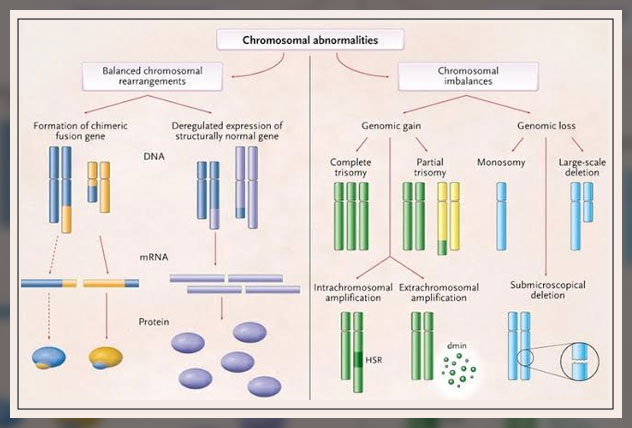
Structural Abnormalities
This occurs when a large section of DNA is missing from or added to a chromosome.
Structural abnormalities can take different forms.
Deletion: A mutation that causes a part of a chromosome to be missing.
Duplication: A mutation causing part of the chromosome to repeat, resulting in extra genetic material.
Translocation: A mutation causing one portion of a chromosome to move to a different part of a chromosome or intrachromosomal.
Or to a different chromosome altogether, i.e. interchromosomal.
There are two key types:
- reciprocal: segments from two different chromosome exchange
- Robertsonian: an entire chromosome attaches to another
Inversion: A mutation resulting in a portion of a chromosome that is in the opposite direction.
Ring: When a portion of the chromosome is broken off and forms a circle or ring.
Learn more about Peripheral Artery Angioplasty Procedure and Stent Placement here.
Scheduling a Scan during Pregnancy
It is important to note that the clear space at the back of the baby can disappear by week 15.
So an NT scan should be completed in the first trimester of the pregnancy.
Moreover, this test can also include blood work to measure the levels of plasma protein and human chorionic gonadotropin, HCG.
HCG is a maternal hormone.
The case of abnormal levels of either can indicate a chromosome problem.
How does NT Scan Works?
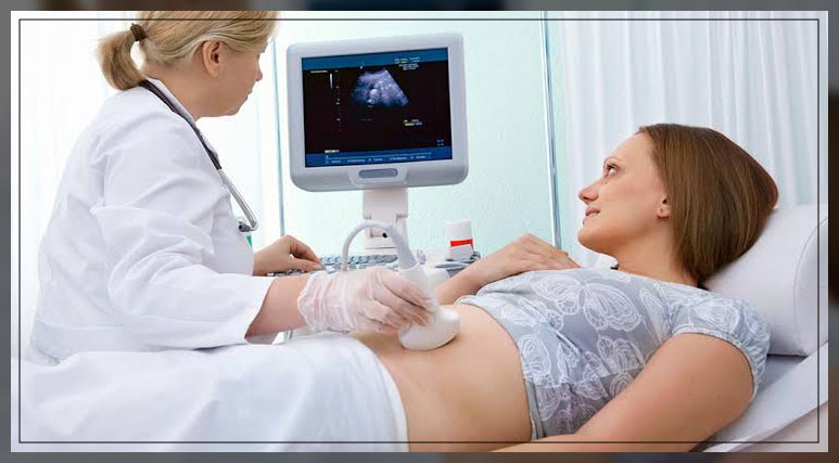
During the screening, the doctor or technician will take an abdominal ultrasound.
The patient alternatively has a transvaginal test as well, where the doctor will insert an ultrasound probe through the vagina.
An ultrasound of high-frequency sound waves creates an image from inside the body of the mother.
From this image, the doctor or technician will measure and carry a nuchal translucency NT scan, translucency or clear space at the back of the neck of the baby.
Moreover, they can also enter the age or date of birth of the patient in a computer program to calculate the risk of the baby having an abnormality.
An NT scan, however, cannot diagnose Down Syndrome or any other chromosome abnormality.
These test only predicts the risk.
Therefore, it is important for the patient to talk to the doctor about available blood tests.
They can also help assess the risk a baby may have.
As with any prediction, the accuracy varies.
If your combine an NT scan with blood testing, the screening tends to be about 85% accurate for predicting the risk of Down Syndrome.
However, if you do not combine blood testing with the scan, the accuracy rate or detection rate drops to about 75%.
Preparing for the Scan
It is important to note that there is nothing special a patient needs to prepare for the NT scan.
In most cases, the test will complete in about 30 minutes.
During the scan, the patient will lie down on an exam table as the technician moves an ultrasound wand over the stomach.
Moreover, ultrasound pictures can be easier to read if the patient has a full bladder, so the doctor can recommend drinking water about one hour before the appointment.
The ultrasound tech will access the lower abdomen of the patient, so it is important for the patient to wear comfortable clothes that make it accessible.
Results from the scan will be available on the same day of testing and the doctor can discuss the finding with the patient before they leave.
It is important to remember that receiving an abnormal result from an NT scan does not necessarily mean that the baby has a chromosome problem.
In the same way, normal test results cannot guarantee that the baby will not be born with Down syndrome.
Furthermore, the test is not perfect. There is a 5% false-positive rate.
In other words, this means that 5% of women can receive positive test results but the baby is fine.
After a positive result, the doctor can suggest another blood test: Prenatal cell-free DNA screening.
This will help examine fetal DNA in the bloodstream of the patient to assess the risk of the baby having Down syndrome and other chromosome abnormalities.
Screening vs. Diagnostic Testing
It is important to note that some patients may find it frightening to receive inconclusive or positive results from an NT scan.
However, keep in mind that an NT Scan can only predict the risk a baby may have.
It does not offer a definitive answer about chromosomal abnormalities.
An NT scan is a screening test, not a diagnostic test.

There are differences between screening and diagnostic testing.
The aim of screening tests is to identify risk factors for a certain disease or condition.
On the other hand, diagnostic testing confirms the presence of a disease or condition.
Diagnosing an Abnormality
In order to diagnose a chromosome abnormality, a patient can ask the doctor about diagnostic testing.
Options include an amniocentesis which is when a doctor will insert a need through the stomach into the amniotic sac to retrieve a fluid sample.
Amniotic fluid contains cells that provide genetic inflammation in the baby.
However, another option is chorionic villus sampling.
A sample of the placental tissue is removed and tested for chromosome abnormalities and genetic problems.
There is a small risk of miscarriage with both tests.
Final Thoughts
An NT scan is a safe, noninvasive test that does not cause any harm to the patient or the baby. Keep in mind, however, that this is a first-trimester screening doctor will recommend but is optional.
NT measurement can help to detect nasal bone, crown rump length, baby’s neck, etc. It can help predict issues before the due date, and high-risk pregnancies. Trisomy 21 helps with a test such as chorionic villus sampling.
Some women may skip this particular test because they do not want to know their risk. Therefore, they can talk to their doctor if they experience anxiety or are worried about how the test results may affect them.