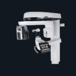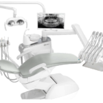
Are you experiencing pain near your molars? Chances are that this is because of an impacted wisdom tooth. In order to check that, get an X-ray at your dentist’s Dental X-ray Unit.
As a dentist yourself, you may be well aware of how important an X-ray is to your setup.
It can give you more clarity about what is exactly happening in your patient’s mouth, clearly showing the teeth and their condition.
Also analyzing the condition of jaws, facial bone composition, and roots.
Different types of X-rays examine your mouth.
Let’s find out below which ones you need for your dental practice or will require to go through if you want a checkup.

What can a Dental X-ray Unit Detect?
X-rays are essentially a form of energy.
It travels through objects or is absorbed.
They can pass through less dense areas such as the gums and cheeks.
Hence, they do not show in the X-ray film but rather appear in darker areas.
On the other hand, teeth and bones are dense objects.
Therefore, they absorb all the X-rays and appear as whiter areas on the film.
A dental X-ray is important to see the oral health problems that are not visible simply by examining your teeth.
In order to get an accurate diagnosis, dentists may recommend patients get an X-ray and confirm their suspicions.
It can also confirm the diagnosis of problems that an oral exam can indicate but a dentist needs further assurity.
These include:
- Tooth decay is present beneath existing fillings and also in small areas between the teeth that are hard to detect. In an X-ray areas with decay appear black. Hence, they are easily detectable in whiter areas of teeth.
- You are showing the condition and position of teeth to have processes such as dental implants, braces, and dentures.
- Detecting abscesses which is an infection between the gum and tooth or the root of the tooth
- A bone loss in the jaw
- Formation of tumors and cysts in the oral cavity
- Identifying changes in root canal or bone as a result of infection.
Dental X-rays are not only for adults.
In fact, children may also need them to confirm their diagnosis and detect bigger issues such as:
- Looking for enough space in the mouth that would fill the coming adult teeth
- The development of wisdom teeth
- The development of tooth decay
- Checking for whether there are impacted teeth that are not unable to erupt properly through gums
There are different types and components of an X-ray unit. Read more below!

What Does a Dental X-ray Unit Entail?
So an X-ray unit consists of 3 main components that further contain equipment:
- X-ray generating equipment which is needed to produce the X-rays
- The image receptors can be in film and digital form in order to detect the X-rays
- Image processing involves using the computer or chemicals to produce the final result which is a black and white image
Let’s talk about these individually in detail.
Dental X-ray Generating Equipment
Every dental X-ray unit price, appearance and complexity may be different.
However, the components are more or less the same.
The three main components include:
- positioning arms
- tube head
- control panel and circuitry
The positioning arm units are usually attached to the wall i.e wall mounted, or floor or can also be attached to a frame on wheels.
However, there are newer handheld dental units as well.
The tube head consists of a glass X-ray tube with a filament, a target, and a copper block.
The transformer provides voltage ranging from 240 Volts to a higher voltage in kV to the X-ray tube.
This is the step-up transformer, whereas the step-down transformer covers the 240 Volts to even lower voltage.
The lead shield minimizes leakage and an oil surrounding it facilitates heat removal.
Additionally, it has a beam indicating device that sets the direction of the beam and the distance from the focal spot to the skin.
A collimator and an aluminum filtration are part of the tube head too.
The control panel consists of the timer, main on and off switch, an exposure time selector, audible signals, and warning lights to control the X-rays that are generated.
There are more components such as the film and the process to produce it.
Let’s dig deeper into that below!

Image Receptors and Processing
For image receptors, you need a radiographic film or digital receptors.
They capture and form the image which is later processed with the help of chemicals and computer technology.
The radiographic film consists of a direct-action film or an indirect-action film.
The direct-action also known as the nonscreen film is used in intraoral radiography.
It is sensitive to X-ray photons.
Hence, it produces an image with fine anatomical detail.
An indirect-action film or a screen film comes in use for extraoral examination.
It includes panoramic radiographs, oblique lateral radiographs, and lateral skull radiographs.

During the process, make sure that the combination of the film and intensifying screen is the same.
Though, different emulsions are sensitive to different light colors that the intensifying screens produce.
The films may contain black paper on either side.
This protects the film from saliva that leaks on the film, light, fingerprints, or other damage by the fingers.
The other way is to get digital images rather than film.
Since there are different types of a dental X-ray machines and their purpose so the result at the end is different too and so are the dental X-ray machine parts.

Types of Dental X-ray Unit
Cone Beam Systems
Using CBCT imaging or Cone Mean Computed Tomography is quickly becoming a standard practice.
It gives 3D imaging that helps in diagnosing oral health problems.
Moreover, the comprehensive view of the anatomy allows for clearer treatment planning.
Intraoral X-ray Sensors
These sensors do not expose the patient to as much radiation as the film X-rays.
In fact, this is one reason why they are quickly replacing film technology.
They allow for quicker and easier tests.
Thus capturing the intraoral images, Full mouth X-ray, and bitewings more efficiently with less exposure.
Phosphor Plate X-ray System
These plates still use film equipment.
It does not include digital imaging however, it uses film equipment with a twist.
These systems involve using reusable plates to capture the high quality images and then feed these plates into a machine that in the end makes them digital.
Next, you have to wipe the plate so that it is clean and ready for use the next time.
However, unlike direct digital imaging devices, these are not as easy to maintain.
They have multiple touchpoints so it is hard to consistently maintain infection control, especially as these will come in use again.
Digital Panoramic X-rays
It uses a small amount of ionizing radiation in order to capture the entire mouth in only one image.
It is a 2D X-ray that takes the upper jaw, lower jaw, tissues, and structures surrounding it in a single image.
The machine also includes additional features like TMJ projections and bitewings.
It helps to streamline diagnosis while also enhancing the practice efficiency.
In fact, some of these are also upgradeable to cone beam imaging and cephalometric.
More on its use is below!

Uses of X-rays in Dentistry
Dental digital radiography such as X-rays can be used for intraoral and extraoral examination.
Intraoral X-rays Include:
- Occlusal X rays
These track the placement and the development of an entire arch.
Hence, it can capture the arch of teeth in the lower and upper jaw.
- Bitewing
It shows upper and lower teeth in a single area of the mouth.
It detects decay and shows the tooth from the crown to the level of the supporting bone as well as changes in the bone thickness due to gum disease.
- Periapical
It shows complete teeth from the crown to further into the root where the teeth attach to the jaw.
It shows all teeth in one portion.
So it is either the upper or the lower jaw.
Moreover, they identify unusual changes in root and bone structures.
Extraoral X-rays
- Tomograms
It shows a layer of the slice of mouth and blurs out all the other layers.
Hence, allowing for more clarity in viewing a particular structure by blurring the rest.
- Sialogram
It uses a dye that is injected into the salivary glands to see them in the X-ray film.
Dentists can ask for this examination in order to check for dry mouth or Sjogren’s syndrome.
- Dental Computed Tomography
It produces an image in 3D.
Its use is primarily in finding problems in the bones such as cysts, fractures, and tumors.
Other types of extraoral X-rays include:
- Cone-beam CT
- Panoramic X-rays
- Cephalometric Projections
- MRI Imaging
- Digital Imaging
Hence, it is an integral part of a dental clinic to detect oral health problems.
Summing Up,
Having a dental X-ray unit within your clinic will allow you to get quicker results for your patients.
This way both diagnosis and treatment will speed up.
Check out these units here to purchase them for your dental clinic setup.


Comments