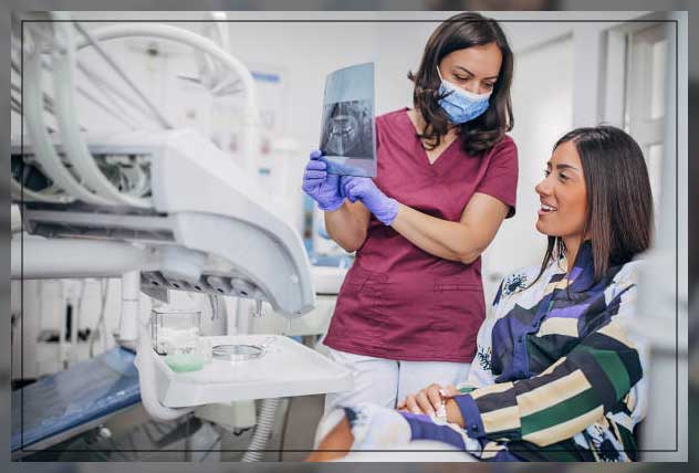
Dental X rays or radiographs are images of your teeth that your dentist will use to evaluate your oral health. These X rays are used with low radiation levels and are hence relatively safe. They capture images of the interior of your teeth and gums. These X-rays can help your dentist identify problems like tooth decay, cavities, impacted teeth, etc.
Though these dental X-rays might seem complex, they are actually widespread tools as vital as your teeth cleanings.
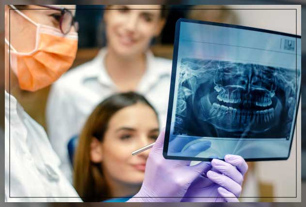
These X-rays help dentists visualize diseases of the teeth, gums, and surrounding tissue that is not visible after a simple oral exam. The X-rays also help them find and treat dental problems early on. This early detection can help save you unnecessary discomfort, money, and even your life.
Read below to learn about dental X-rays and how they are done.
Why Dental X Rays Performed?
Dental X rays are typically performed yearly. They might be done more often if your dentist is tracking the progress of your dental problem or treatment.

Factors that affect how often you get dental X rays might include:
- your current oral health
- your age
- any symptoms of oral disease
- a history of gum disease, gingivitis, or tooth decay
If you are a new patient, you will probably undergo dental X-rays. The reason being that your dentist can get a clear picture of your dental health. This is especially important if you do not have any X-rays reports from your previous dentist.
Children and infants might need to have dental X-rays more often than adults. It is probably because their dentists may need to monitor the growth of their permanent or adult teeth. This is crucial because it can help the dentist determine if baby teeth or milk teeth need to be pulled to prevent all kinds of complications. It is important to know that adult teeth grow behind baby teeth.
Current Technology on Dental X Ray
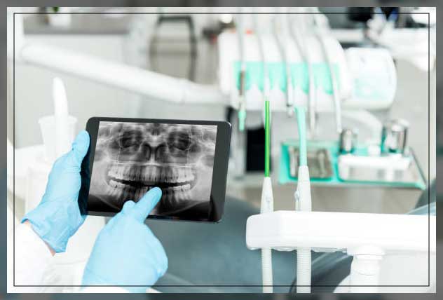
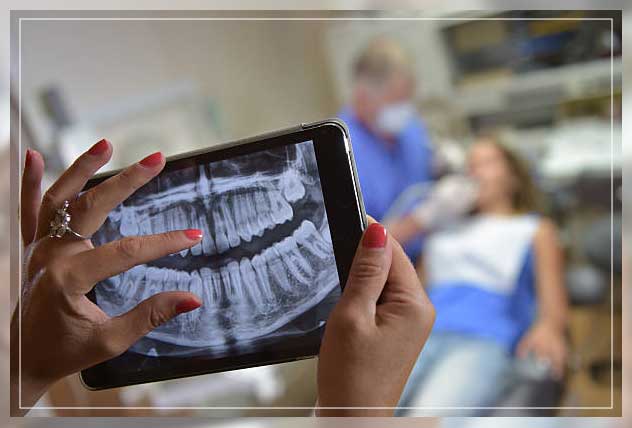
There is a newer dental X-ray technique most dentists use today. Your dentist already might be using or may soon be using. It is called digital imaging.
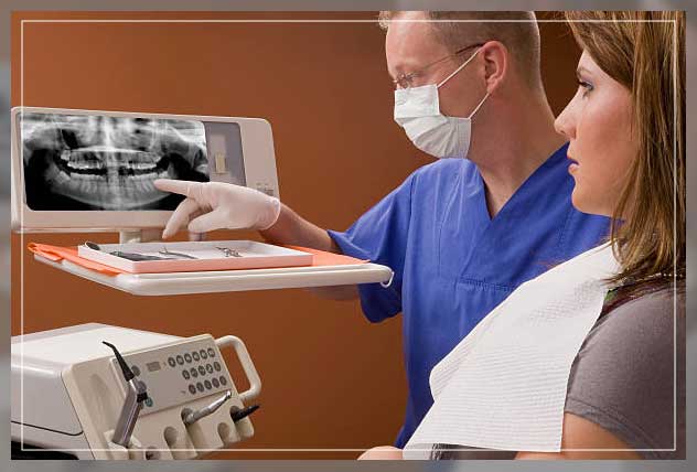
Instead of developing X-ray film in a dark room, your dentist will send these X-rays directly to a computer. Then they can view them on-screen, stored, or print them out.
Moreover, there are several benefits of using this new technology:
1 This new technique uses less radiation than the typical X-ray. Moreover, there is no wait time for the X-rays to develop. The images are available on the screen a few seconds after being taken.
2 The image of a tooth or gum can be enhanced and enlarged several times. This enlarged image on the computer screen makes it easier for your dentist to show you where and what the problem is.
3 Another advantage is that the images can be electronically sent to another specialist or dentist – for instance, for a second opinion on a dental problem. It is often done to determine if a specialist is needed for the treatment.
4 Software added to the computer can help your dentist digitally compare current images to previous ones. This process is known as subtraction radiography. Using this technique, everything that is the same between the two images is “subtracted out”. Therefore, it leaves a clear image of only the portion that is different. Your dentist can easily see the tiniest changes that the naked eye might not have noticed using this technique.
What Problems Can Dental X-Rays Detect?
In adults, X-rays can be used to:
- Identify areas of decay that might not be visible with an oral exam, especially small areas of decay between teeth.
- Identify decay occurring beneath an existing filling.
- Reveal bone loss that might accompany gum disease.
- Reveal changes in the bone or in the root canal that might result from an infection.
- Assist in preparing tooth implants, dentures, braces, or other procedures.
- Reveal an abscess, an infection occurring at the root of a tooth or between the gum and a tooth.
In children, X-rays are used to:
- Watch for decay in their milk or permanent teeth.
- Determine if there is enough space in the mouth to fit all incoming teeth.
- Determine if primary teeth are being lost at the correct time to allow permanent teeth to come in properly
- Check for the development of wisdom teeth. Also, identify if the teeth are impacted – teeth that cannot emerge through the gums.
- Reveal other developmental abnormalities, such as cysts and some types of tumors that may occur.
Risks of Dental X-rays
While dental X rays do involve radiation, the exposure levels are so low that they are considered safe for adults and children. If your dentist uses digital X-rays instead of developing them on film, then your risks from radiation exposure are even lower. As they are safer.

Your dentist will also place a lead bib just over your abdomen, chest, and pelvic region. This lead bib will help prevent any unnecessary radiation exposure to your vital organs. Your dentist may also use a thyroid collar in the case of thyroid conditions. Kids and women of childbearing age might also wear them along with the lead jacket. Your dentist may use other dental instrument as well.
Note that pregnancy is an exception to the rule. Pregnant women or women who believe they might be pregnant should avoid all types of X rays. Tell your dentist if you think you are pregnant, as the radiation is not considered safe for your developing fetuses.
Preparing for Dental X Ray
You should be aware that dental X rays require no special preparation. The only important thing is to brush your teeth before your appointment. It will create a more hygienic, clean env for those working inside your mouth.
You will sit in a chair with a lead vest across your chest and abdomen at the dentist’s office. Your specialist positions the X ray machine alongside your head to record images of your mouth. It is one of the important lap equipment you dentist might use.
In some dental clinics, they have a separate room for X-rays. While others perform them in the same room as cleanings and other procedures are carried out.
Types of Dental X Ray
There are several types of dental X-rays. They are used to record slightly different views of your mouth. The most common ones are intraoral X-rays.
Bitewing. Your dentist will use this technique – when you bite down on a special piece of paper, and your dentist will check how well the crowns of your teeth match up. This technique is commonly used to check for tooth cavities between teeth (interdental). As these cavities are not visible easily.
Occlusal. Your dentist will use this technique as it captures all of your teeth in one shot.
Occlusal. Your dentist will opt for this X-ray when your jaw is closed to see how your upper and bottom teeth line up. It can also successfully detect anatomical abnormalities with the palate or floor of your mouth.
Panoramic. For this type of X-ray, the machine rotates around the head. Your dentist may use this technique to check your wisdom teeth, plan for implanted dental devices, or investigate jaw problems.
Periapical. This technique focuses on two complete teeth from root to crown.
Extraoral. X-rays may be used when your dentist suspects there might be problems in areas outside of the gums and teeth, such as the jaw.
Your dentist will guide you through each step of the X-ray process. They may step outside the room briefly while the machine takes your images.
Your dentist will ask you to hold still while the pictures are recorded. In case they are used, Spacers or film holders will be moved and adjusted in your mouth to obtain the proper images.
After a Dental X Ray
Once the images are ready, your dentist will review them. It is only possible in the case of digital X-rays. The images are ready instantly. Your dentist will examine them and check for any abnormality.

If your dentist finds problems or identifies issues, such as tooth cavities or tooth decay, they will discuss with you the treatment options. If your dentist finds no problems, then you can go home happily.
How Often Should Teeth Be X-Rayed?
The frequency depends on your medical and dental history and your current condition. Some people might need X-rays as often as every six months. In contrast, others with no recent dental or gum disease and who visit their dentist regularly may get X-rays only every couple of years.
However, if you are a new patient, your dentist might take X-rays as part of the initial exam. It would help establish a baseline record to compare the dental changes that might occur over time.
The Outlook
Like brushing and flossing, getting a regular dental X-ray when you go for a scheduled check is also essential. Note that it is an integral part of your overall oral health.
Hope you would agree that having a good checkup can be a relief. But it does not mean you should not keep getting X-rays.
Depending on your age, health, and insurance coverage, your dentist may perform X-rays once every one to two years. Be sure to commit to your appointments and visit your dentist sooner if you experience any pain or other changes in your mouth.


Comments