
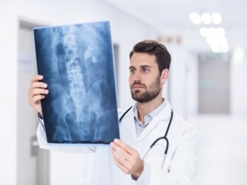
This is probably one of the most important medical equipment to know of. X-ray machine falls under the category of radiography.
It is what we consider as a medical imaging technique. And it revolves around the need to take images of the structures inside the body.
And these are the images which will be viewed on a digital platform.
These X-rays are important for a range of reasons.
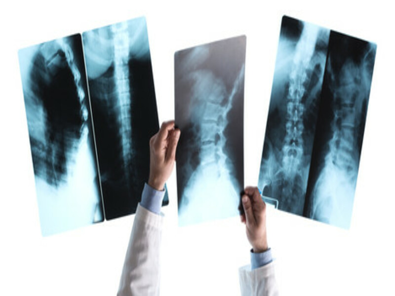
You will be needing them to look into the bones and teeth. So it focuses on helping in the diagnosis of different fractures. Including that of broken bones.
As well as certain diseases called as arthritis. So a healthcare provider is going to look into and determine all there is to know regarding the structures of the chest, including that of lungs, heart, breasts and more body parts.
In this article I will be looking into and speaking in detail what is it that x-ray imaging is. How it works, the different types and more.
So let’s begin:
What Are Medical X-rays?
This is what we consider as being electromagnetic radiation. A medical imaging technique which is like that of visible light.
However when looking into X-rays, they will be having more energy which means they can easily pass through various objects.
Including that of passing through the body as well. These kinds of X-rays are important as they will be generating different kinds of tissues and structures within the body.
Also know that if an X-ray is going to be passing through the X-ray detector, an image will be forming which is representing what we call as the shadows.
When we speak of electromagnetic radiation, this is a kind which is traveling like waves. And it also consists of electric as well as magnetic fields.
Looking at the devices which make use of this kind of radiation include infrared light, microwaves, gamma rays, visible light and of course X-rays.
How Do They Work?
Now lets look into how do they even work?
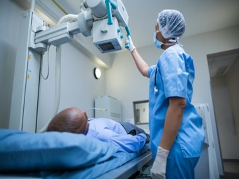
First of the patient is made to position in a way where the part of the body which needs an X-ray imaging is coming in between the X-ray machine and the X-ray detector.
So when this machine is turned on, the X-rays will be travelling through the body. And when they do, it is absorbed in different amounts.
This is through the tissues of the body. And also it directly depends on the radiological density of the tissues.
What is radiological density?
It is determined from not just the density but also the atomic number which is of the materials being imaged.
Remember that bone structures consist of calcium. This is something which has a high atomic number as compared to other tissues.
This is probably why bone structures actually appear to be whiter in appearance. The tissues are actually looking far more whiter against the black background of the radiograph.
And due to this property, bones will be able to readily absorb the x-rays which travel through the body.
Remember one thing – the X-rays will be travelling far more easily through this path field as compared to other such medical imaging tools. They also travel faster through the less dense tissues like fat and muscle.
You will notice how these structures are getting displayed through the shades of gray.
Now let’s talk about where do we use these?
The Use of X-rays and X-ray Machine
Now that we know of what is an X-ray machine and how it creates X-rays, lets now look into and talk of ways in which we can adapt its use and see results.
There are plenty of ways in which medical experts will be using x-ray machine to get solid results.
That means understanding how it will be emphasizing X-ray machines have so many ways of creating images that are going to bring forth a way of diagnosing the concerns and how it will be yielding strong imaging.
Diagnostic Uses
When we look at how x-ray radiography works, it basically aims at detecting the bone fractures, as well as certain tumors. Also it will be looking at the abnormal masses, pneumonia issues as well as certain injuries that may be taking on.
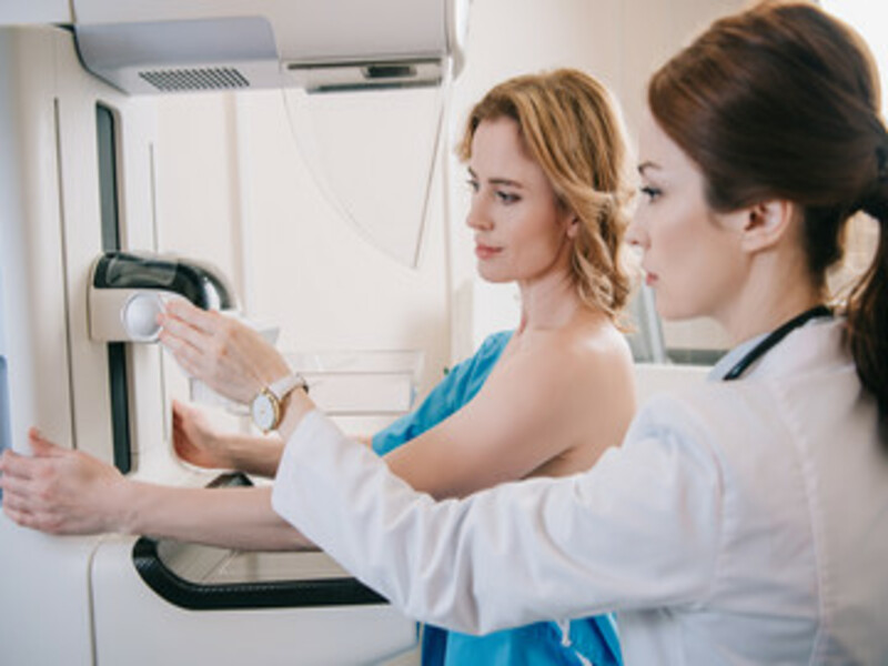
Another important use of this medical imaging is for mammography purposes. It is essential for detecting breast cancer.
It is a solid way of early cancer detection.
How?
Because the tumor is going to be appearing as either regular or irregular shaped mass. And this comes brighter in the imaging with the black background.
Know this- mammograms are also solid ways of detecting calcium called microcalcifications.
They appear as really small, tiny specks against the screen.
Use in Computed Tomography
Another important use of x-ray imaging is when we use it in CT. This is the combination of traditional x-ray technology with the computer processing.
It will be generating a range and series of cross-sectional images.
You will be combining them with three-dimensional x-ray imaging to get solid results.
CT images are actually more in use.
They will be providing you with more details as compared to plain radiographs.
Also they work great as they give you the ability to view the structures in a more broader and wider angle.
That will in itself confirm how this imaging is yielding solid results.
Fluoroscopy
I also want to share details on how you will be making use of x-rays as well as a fluorescent screen. This is done so you can obtain real-time images. They aim at movement within the body and help with the diagnostic process.
It includes ways in which you will be seeing how you are basically injecting some kind of radiographic contrast agents.
That will suggest and provide an overview as to how the blood is flowing to the heart muscle as well as all the different blood vessels.
Many prefer this kind of diagnosis for two main reasons- one of that it is internally threaded and you will be getting to see results in a simple and fast manner.
Secondly it basically relates to how it is a minimal invasive procedure. And you aim for the same to open up clogged arteries, which are responsible for supplying blood to the heart.
Does It Have Therapeutic Use?
So this is something many may ask of.
How does it hold certain therapeutic measures?
See radiation therapy basically relates to how in cancer treatment, we will be making use of X-rays as well as other types of high-energy radiation which we can use to actually destroy the cancer tumors.
But what is the basis of its application?
The radiation dose is used in small amounts. It is smaller as compared to the one used in diagnostic imaging.
The therapeutic radiation is such that it will be coming from a machine outside of the body.
Or from the radioactive material as well which is used in the body, or inside near the tumor cells. Most probably being injected right into the blood stream as well.
X-ray Machine Risks
It is important to identify how we can safely make use of this method. There are definitely certain risks associating with it.
While this is like a life-saving process.
When it comes to diagnosing very serious illnesses like cancer, infections and other ailments, it is important to know and consider the minimal risks that come with this process.
One thing to know is that X-rays actually produce an ionizing radiation. This is a form of radiation which will be harming the living tissue.
Also the risks may increases as the exposure increases to life.
But know one thing- the risks of developing cancer from such radiation exposure is very small.
Yet for a pregnant woman, getting an x-ray image for the pelvis or abdomen can pose serious problems.
Similarly in children, they are definitely more sensitive to the ionizing radiation. And this is why it serves with a higher risk of developing cancer than in adults.
Yet, this number falls into a smaller circle of possibility.
Overall, X-ray machine is used all over the world for many reasons. It is definitely an essential element for diagnosing illnesses hence you have to see the advantages of it over the disadvantage.
Takeaway
When we look into and speak of certain medical equipment, we need to see how effective is its use. There are so many ways in which it will be leading to providing essential information for your medical healthcare provider.
Among the many kinds, X-ray machine is essentially one of the most important ones.
It relates to how you will be handling the diagnosis process by making use of medical imaging.
In this article we have spoken at great lengths to what is an X-ray machine, the kind of images it creates. And how we can make use of them.
All of these important details emphasize why we should be adapting this technology .
Certain risk factors were also looked at however it is certain that there are far more uses of this machine which makes it an essential tool for the world of medicine.


Comments